Basic HTML Version


Chapter 3 — 3
Below the larynx the air passes through a tube called the
trachea
. This is about as thick as the
average snorkel and branches inside the chest into two tubes, the
bronchi
, which lead to the
lungs. Those air passages are lined with cells covered with microscopic hairs (cilia) which move
a sheet of secreted mucous slowly upwards towards the larynx. Small pieces of foreign material
such as dust eventually find their way to the larynx, along with this mucous sheet. It is then
either coughed-up or swallowed. The cilia may be damaged by smoking or infection, causing
retention of mucous and inhaled material which may eventually obstruct the air passages.
The bronchi divide repeatedly into progressively smaller passages rather like the branches of a
tree. These passages have encircling muscles in their walls which, by contraction or relaxation,
can vary the diameter of the air passage.
In
asthma
the muscles of the small bronchi become oversensitive and overactive, causing
excessive narrowing and obstruction of these air passages. This can occur in response to
exercise, allergy, cold, infection, anxiety, smoking or other inhalants such as sea water. At the
same time, the cells lining these passages produce excessive and thickened mucous. The
combination of these factors causes airway narrowing which has serious repercussions for a
diver.
The smallest branches of the bronchi end in bunches of microscopic air sacs called
alveoli
. The
vast number of alveoli are packed together into the two sponge like organs, the
lungs
. There are
about 300 million alveoli in the lungs and the combined surface area of all the alveoli in the
lungs is equal to about half a tennis court. The alveoli are lined by a thin layer of fluid containing
a detergent-like substance called
surfactant
. This acts as a wetting agent to prevent the alveoli
from collapsing from surface tension.
The surfactant lining of the alveoli can be damaged in disease or by inhalation of water, leading
to collapse of the lungs and serious respiratory difficulty.
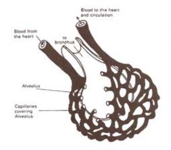
Each alveolus is surrounded by a network of blood capillaries. These bring the blood into close
contact with the air in the alveolus, with only the microscopically thin walls of the alveolus and
capillary separating the two.
Fig. 3.2
This diagram illustrates an alveolus with its surrounding meshwork of
capillaries.